


When Ph.D. student Blake Rogers talks about microscopes, he sounds like a kid on Christmas morning. The enthusiasm is real, and rightly so. The microscope of which he speaks is not just any microscope, but a transmission electron microscope (TEM).
Most microscopes, common in labs, are limited to magnifications of about 1,500 times.
The TEM can easily achieve magnifications of up to two million times. And NAU acquired a new one. (Hence, his excitement.)
 Rogers had a successful career before deciding to pursue his doctorate. He worked for Intel, designing and testing thermal cooling solutions for high-performance processor packages. Then, he designed and fabricated test reactors for PET recycling at a startup company. But he was ready for a change.
He had heard good things about NAU’s newly created Department of Applied Physics and Materials Science (APMS) and wanted to be involved. He didn’t know, however, that part of that involvement meant helping NAU acquire a new TEM.
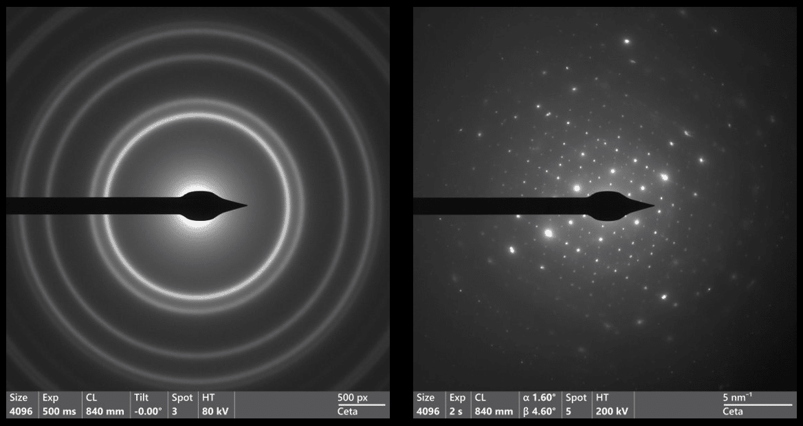
“The transmission electron microscope is a fantastic instrument for scientific research,” Rogers said. “The most exciting capabilities of the TEM are its ability to resolve features on an atomic scale, the ability to quickly switch between imaging and diffraction studies (more accurate and much more informative than imaging) and the ability to map out the elements in a material in detail.”
The last time NAU purchased one of these microscopes was in the early ‘80s, and while it is still operational, the newer TEM can produce much higher magnifications.
“Not only can this TEM resolve individual columns of atoms, but in EDX mode it can identify the atoms by characteristic X-ray emissions allowing a group to map out the positions of various elements in a sample. All of this combines to provide NAU students with the ability to conduct research at the highest levels in science.”
Currently, several groups on campus are using it for materials research. Professor Miguel José Yacamán, a world-renowned TEM expert who was the driving force behind obtaining the machine, leads a group of students studying nanoparticles of many shapes, sizes and compositions. Other groups are using it to understand the process of synthesizing light activated nano-jets that can be directed to swim through liquids in specific directions. Researchers at W. L. Gore have been using it to characterize cellulose fibers that were bio-engineered. And Rogers? He’s taking blurry images, on purpose.
Together with APMS professor Chris Mann, Rogers is helping develop methods for in-line electron holography or phase retrieval. By analyzing a series of images that are progressively more out of focus, they can reconstruct the sample wave function and generate images that are not only in focus, but also corrected for aberrations inherent to the TEM, specifically for fragile samples like muscle tissue. And when he isn’t taking images out of focus, he is studying samples using diffraction techniques, some of which are also performed out of focus.
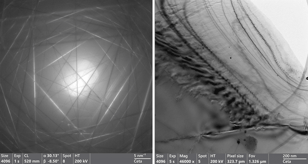
“One of the coolest things I have seen is a convergent beam diffraction pattern of a nanoparticle with five-fold symmetry,” Rogers said. “This is cool because in most crystal systems, five-fold symmetry is geometrically impossible, but nanoparticles are so small that it can and does exist on that level. And the TEM allows us to see it.”
Any group on or off campus can be trained to use the microscope. The internal rate is $50 per hour, and the external rate is $150 per hour. To inquire about TEM training and use, email Joanna Stephens.
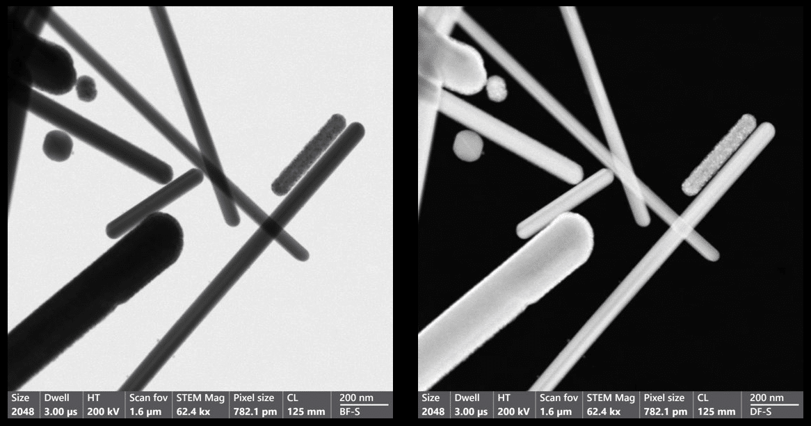
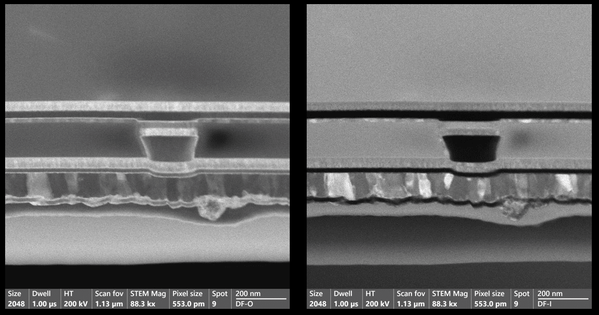
Check out some of the images capture by the TEM below.
 Carly Banks | NAU Communications
(928) 523-5582 | carly.banks@nau.edu